TORONTO — Cellphones with built-in cameras have been a real boon for the YouTube set or anyone who wants to capture a spur-of-the-moment photo. Now researchers have a new use for the pocket-sized gizmos — as a means of capturing microscopic data for diagnosing disease.
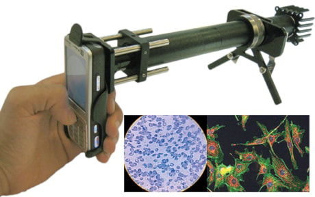
Biomedical engineers at the University of California, Berkeley, have developed a prototype camera-phone mounted with a microscope to magnify and photograph blood or saliva samples for testing.
The prototype CellScope, as it’s been dubbed, would allow disease screening and diagnosis in the field where specialized clinical microscopy laboratories aren’t available, including in underdeveloped countries or isolated or rural locations in the developed world.
“The same regions of the world that lack access to adequate health facilities are, paradoxically, well-served by mobile phone networks,” said Dan Fletcher, an associate professor of bioengineering and head of the team developing the CellScope.
“We can take advantage of these mobile networks to bring low-cost, easy-to-use lab equipment out to more remote settings.”
David Breslauer, a graduate student on the team who specializes in developing small medical devices, said there is increasing interest in expanding mobile technology for medicine, whether for monitoring a person’s heartbeat or checking a diabetic’s blood glucose levels.
“And for many diseases, simply looking through a microscope is still the standard,” Breslauer said Tuesday from Berkeley, Calif. “It’s not like a pregnancy test, where it says yes or no. You look through a microscope, a trained professional sits there and counts whatever they’re looking for and reports it.”
Cellphone cameras have already been used to snap shots of magnified blood samples, for instance, by holding the device up to the eyepiece of a microscope, he said.
“But what we wanted to do was actually design a microscope around the cellphone, make a portable system that could then be used to do clinical-level diagnostics that require light microscopy in a portable field-ready fashion.”
In testing of samples of infected blood and saliva, the researchers were able to use CellScope to capture pictures of the parasite that causes malaria as well as fluorescent-tagged images of the skinny rod-like bacteria that leads to tuberculosis.
“The images can either be analyzed on site or wirelessly transmitted to clinical centres for remote diagnosis,” said Breslauer, co-lead author of a report on the device published this week by the journal PLoS One.
“The system could be used to help provide early warning of outbreaks by shortening the time needed to screen, diagnose and treat infectious diseases.”
Breslauer said the group is working on a more robust version of CellScope — the current prototype is quite fragile and would likely break if dropped — that can be taken out in the field for testing in countries like Honduras, the Democratic Republic of Congo and Tanzania.
With under $75 in parts, Dan Fletcher, UC Berkeley associate professor of bioengineering and head of the research team developing the CellScope demonstrated the feasibility of the first generation CellScope.
Next, they will develop, test, and deploy a second-generation CellScope with an even greater magnification and with both transmitted and reflected light illumination that has the potential to enhance access to specialty health services.
The idea would eventually be to equip travelling health providers with the devices, so that people living long distances from hospitals or clinics could be tested for diseases without having to leave home. They could also be used for post-treatment follow-up.
“If you can give someone this system, then they can go from house to house,” he said. “And because the cellphone is a computer essentially, they can input patient data, they can geotag their location with a GPS (global positioning system), and so you shift where the health care’s being provided.”
In developed countries like the United States and Canada, the microscope camera could be used by a cancer patient having chemotherapy, for example, to take regular snaps of their blood.
The captured images could be sent by home computer to a physician, who could remotely monitor the patient’s white blood-cell count.